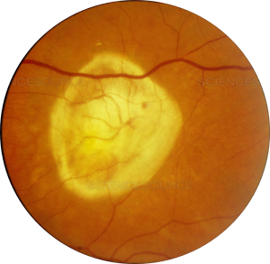
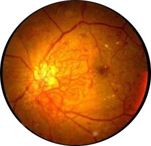
 1. How does the eye work?
When you take a picture with a camera, the lens in the front of the camera allows light to pass through and focus that light on the film that covers the back side of the camera. A picture is taken when the light hits the film. Our eyes work in a very similar way. The front of the eye (the cornea, pupil and lens) is clear, which allows light to pass through. The cornea and lens of the eye focuses the light on the back wall of the eye, the retina. Like the film, the retina is the “seeing” tissue of the eye, sending messages to the brain through the optic nerve, allowing us to see.
2. When should an adult’s eyes be examined?
We recommend adult examinations of the eyes be performed on a regular basis. Below is a chart with a recommended time line of how often an adult should receive an eye examination.
Ages 20-39 Every three to five years.
Ages 40-65 Every two to four years.
Ages 65 and older Every one to two years.
3. What is a Vitreo-retinal specialist?
Vitreo-retinal specialists treat eye diseases that affect the back of the eye, such as retinal detachment, macular degeneration, and diabetic retinopathy. Our eye physicians and surgeons have received a medical degree and completed a residency in ophthalmology and a retina fellowship to provide patients the highest quality eye care.
4. What is the vitreous?
The vitreous a thick gel-like substance that fills most (80%) of the eye. It helps maintain the round shape of the eye. It contains fibers that are attached to the retina.
5. What is retinal detachment?
Retinal detachment occurs when the retina separates or pulls away from the back of the eye. This can happen when vitreous liquid seeps through a small tear or hole in the retina and builds up under the retina. As vitreous liquid accumulates under the retina, it can detach from the blood vessels that provide oxygen and nutrients. The risk of permanent vision loss increases as the detachment remains untreated. Floaters are a common symptom of retinal detachment.
 6. What are floaters?
Floaters are small specks of debris that move in and out of your field of vision.
7. Who are at risk for retinal detachments?
People over 40 are more likely to experience retinal detachment. Previous retinal detachment, a family history of retinal detachment, and extreme nearsightedness can also put you at a higher risk.
8. How are vitreo-retinal problems treated?
Retinal tears and detachment, eye disease and trauma can cause vision loss. Fortunately, surgery and lasers can be used to treat most conditions before vision gets worse.
9. What is diabetic retinopathy?
Diabetic retinopathy is an eye disease that damages the blood vessels in the retina. People with Type I and Type II diabetes can experience diabetic retinopathy. In advanced stages, diabetic retinopathy can cause blurry vision, floaters, vision loss and ultimately blindness. Diabetics can prevent diabetic retinopathy by managing their blood pressure, cholesterol and blood sugar.
10. What is Age-Related Macular Degeneration?
Age-Related Macular degeneration (AMD) is a breakdown of the macula, which is located in the center of the retina, and provides sharp central vision that is necessary for reading, driving and other important tasks. There are two forms of AMD: Wet and Dry. Dry AMD is more common. With this condition, the macula deteriorates, causing blurry vision. Wet AMD is more damaging and can lead to serious vision loss.
